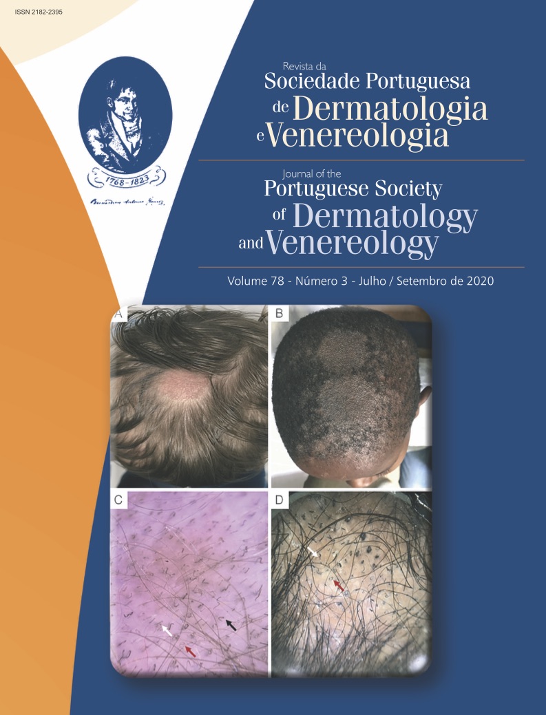
Dermoscopy in Pediatric Dermatology – Part II: Infectious and Inflammatory Skin Lesions
Abstract
Dermoscopy is a noninvasive technique that increases diagnostic accuracy of several skin lesions. Being painless, dermoscopy is particularly useful in children, sparing them from unnecessary biopsies and treatments. In part II of this article, we highlight the importance of dermoscopy for the diagnosis and follow-up of infectious and inflammatory skin disorders in pediatric patients.
Downloads
References
Haliasos EC, Kerner M, Jaimes N, Rudnicka L, Zalaudek I, Malvehy J, et al. Dermoscopy for the Pediatric Dermatologist Part I: Dermoscopy of Pediatric Infectious and Inflammatory Skin Lesions and Hair Disorders. Pediatr Dermatol. 2013;30(2):163-71. doi: 10.1111/pde.12097.
Micali G, Giuffrida G, Quattrocchi E, Lacarrubba F. Scabies. In: Micali G, Lacarrubba F, Stinco G, Argenziano G, Neri I, editors. Atlas of Pediatric Dermatoscopy. Geneve: Springer; 2018. p.53-61.
Hill TA, Cohen B. Scabies in babies. Pediatr Dermatol. 2017;34(6):690–694. doi:10.1111/pde.13255.
Zalaudek I, Giacomel J, Cabo H, Di Stefani A, Ferrara G, Hofmann-Wellenhof R, et al. Entodermoscopy: a new tool for diagnosing skin infections and infestations. Dermatology. 2008;216(1):14–23. doi:10.1159/000109353.
Suh KS, Han SH, Lee KH, Park JB, Jung SM, Kim ST, et al. Mites and burrows are frequently found in nodular scabies by dermoscopy and histopathology. J Am Acad Dermatol. 2014;71(5):1022–1023. doi:10.1016/j.jaad.2014.06.028.
Neri I, Chessa MA, Virdi A, Patrizi A. Nodular scabies in infants: dermoscopic examination may avoid a diagnostic pitfall. J Eur Acad Dermatol Venereol. 2017;31(12):e530-e531. doi: 10.1111/jdv.14401.
Chavez-Alvarez S, Villarreal-Martinez A, Argenziano G, Ancer-Arellano J, Ocampo-Candiani J. Noodle pattern: a new dermoscopic pattern for crusted scabies (Norwegian scabies). J Eur Acad Dermatol Venereol. 2018;32(2):e46–e47. doi:10.1111/jdv.14498.
Micali G, Lacarrubba F, Verzì AE, Chosidow O, Schwartz RA. Scabies: Advances in Noninvasive Diagnosis. PLoS Negl Trop Dis. 2016;10(6):e0004691. doi: 10.1371/journal.pntd.0004691.
Rubegni P, Mandato F, Risulo M, Fimiani M. Non-invasive diagnosis of nodular scabies: the string of pearls sign. Australas J Dermatol. 2011;52(1):79. doi:10.1111/j.1440-0960.2010.00686.x.
Lacarrubba F, Boscaglia S, Dinotta F, Micali G. Pediculosis. In: Micali G, Lacarrubba F, Stinco G, Argenziano G, Neri I, editors. Atlas of Pediatric Dermatoscopy. Geneve: Springer; 2018. p.63-70.
Del Giudice P, Hakimi S, Vandenbos F, Magana C, Hubiche T. Autochthonous Cutaneous Larva Migrans in France and Europe. Acta Derm Venereol. 2019;99(9):805–808. doi:10.2340/00015555-3217.
Aljasser MI, Lui H, Zeng H, Zhou Y. Dermoscopy and near-infrared fluorescence imaging of cutaneous larva migrans. Photodermatol Photoimmunol Photomed. 2013;29(6):337–338. doi:10.1111/phpp.12078.
Piccolo V. Update on Dermoscopy and Infectious Skin Diseases. Dermatol Pract Concept. 2019;10(1):e2020003. doi:10.5826/dpc.1001a03.
Grayson W. Infectious diseases of the skin. In: Calonje JE, Brenn T, Lazar A, Billings S, editors. McKee's Pathology of the Skin: with Clinical Correlations, 5th edn. Philadelphia: Elsevier Saunders; 2019. p.972.
Pimenta R, Soares-de-Almeida L, Arzberger E, Ferreira J, Leal-Filipe P, Mendes-Bastos, et al. (in press). Reflectance confocal microscopy for the diagnosis of skin infections and infestations. Dermatol Online J.
Gupta AK, Mays RR, Versteeg SG, Piraccini BM, Shear NH, Piguet V, et al. Tinea capitis in children: a systematic review of management. J Eur Acad Dermatol Venereol. 2018;32(12):2264–2274. doi:10.1111/jdv.15088.
Lacarrubba F, Boscaglia S, Micali G. Tinea Capitis. In: Micali G, Lacarrubba F, Stinco G, Argenziano G, Neri I, editors. Atlas of Pediatric Dermatoscopy. Geneve: Springer; 2018. p.45-51.
Lencastre A, Tosti A. Role of trichoscopy in children's scalp and hair disorders. Pediatr Dermatol. 2013;30(6):674–682. doi:10.1111/pde.12173.
Lacarrubba F, Verzì AE, Dinotta F, Scavo S, Micali G. Dermatoscopy in inflammatory and infectious skin disorders. G Ital Dermatol Venereol. 2015;150(5):521–531.
Brasileiro A, Campos S, Cabete J, Galhardas C, Lencastre A, Serrão V. Trichoscopy as an additional tool for the differential diagnosis of tinea capitis: a prospective clinical study. Br J Dermatol. 2016;175(1):208‐209. doi:10.1111/bjd.14413.
Errichetti E, Stinco G. Dermoscopy in General Dermatology: A Practical Overview. Dermatol Ther (Heidelb). 2016;6(4):471–507. doi:10.1007/s13555-016-0141-6.
Lacarrubba F, Verzì AE, Quattrocchi E, Micali G. Cutaneous and Anogenital Warts. In: Micali G, Lacarrubba F, Stinco G, Argenziano G, Neri I, editors. Atlas of Pediatric Dermatoscopy. Geneve: Springer; 2018. p.35-44.
Costa-Silva M, Azevedo F, Lisboa C. Anogenital warts in children: Analysis of a cohort of 34 prepubertal children. Pediatr Dermatol. 2018;35(5):e325–e327. doi:10.1111/pde.13543.
Morais RB, Valério M, Amaro C. Verrugas anogenitais na criança – um desafio diagnóstico. J Port Soc Dermatol Venereol. 2015;73(1):97-104. https://doi.org/10.29021/spdv.73.1.348
Li X, Yu J, Thomas S, Lee K, Soyer HP. Clinical and dermoscopic features of common warts. J Eur Acad Dermatol Venereol. 2017;31(7):e308–e310. doi:10.1111/jdv.14093.
Dong H, Shu D, Campbell TM, Frühauf J, Soyer HP, Hofmann-Wellenhof R. Dermatoscopy of genital warts. J Am Acad Dermatol. 2011;64(5):859–864. doi: 10.1016/j.jaad.2010.03.028.
Lacarrubba F, Verzì AE, Dinotta F, Micali G. Molluscum Contagiosum. In: Micali G, Lacarrubba F, Stinco G, Argenziano G, Neri I, editors. Atlas of Pediatric Dermatoscopy. Geneve: Springer; 2018. p.27-33.
Meza-Romero R, Navarrete-Dechent C, Downey C. Molluscum contagiosum: an update and review of new perspectives in etiology, diagnosis, and treatment. Clin Cosmet Investig Dermatol. 2019;12:373–381. doi:10.2147/CCID.S187224.
Ianhez M, Cestari Sda C, Enokihara MY, Seize MB. Dermoscopic patterns of molluscum contagiosum: a study of 211 lesions confirmed by histopathology. An Bras Dermatol. 2011;86(1):74–79. doi:10.1590/s0365-05962011000100009.
Ku SH, Cho EB, Park EJ, Kim KH, Kim KJ. Dermoscopic features of molluscum contagiosum based on white structures and their correlation with histopathological findings. Clin Exp Dermatol. 2015;40(2):208–210. doi:10.1111/ced.12444.
Micali G, Boscaglia S, Musumeci ML, Lacarrubba F. Psoriasis. In: Micali G, Lacarrubba F, Stinco G, Argenziano G, Neri I, editors. Atlas of Pediatric Dermatoscopy. Geneve: Springer; 2018. p.79-85.
Musumeci ML, Lacarrubba F, Verzì AE, Micali G. Evaluation of the vascular pattern in psoriatic plaques in children using videodermatoscopy: an open comparative study. Pediatr Dermatol. 2014;31(5):570–574. doi:10.1111/pde.12283.
Golińska J, Sar-Pomian M, Rudnicka L. Dermoscopic features of psoriasis of the skin, scalp and nails - a systematic review. J Eur Acad Dermatol Venereol. 2019;33(4):648–660. doi:10.1111/jdv.15344.
Farias DC, Tosti A, Chiacchio ND, Hirata SH. Aspectos dermatoscópicos na psoríase ungueal [Dermoscopy in nail psoriasis]. An Bras Dermatol. 2010;85(1):101–103. doi:10.1590/s0365-05962010000100017.
Piraccini BM, Starace M. Nail Disorders. In: Micali G, Lacarrubba F, Stinco G, Argenziano G, Neri I, editors. Atlas of Pediatric Dermatoscopy. Geneve: Springer; 2018. p.178-179.
Errichetti E, Stinco G. Lichen Planus. In: Micali G, Lacarrubba F, Stinco G, Argenziano G, Neri I, editors. Atlas of Pediatric Dermatoscopy. Geneve: Springer; 2018. p.87-93.
Pandhi D, Singal A, Bhattacharya SN. Lichen planus in childhood: a series of 316 patients. Pediatr Dermatol. 2014;31(1):59–67. doi:10.1111/pde.12155.
Payette MJ, Weston G, Humphrey S, Yu J, Holland KE. Lichen planus and other lichenoid dermatoses: Kids are not just little people. Clin Dermatol. 2015;33(6):631–643. doi:10.1016/j.clindermatol.2015.09.006.
Oliveira A, Mendes-Bastos P. Multiple annular plaques on a 9-year-old boy. Pediatr Dermatol. 2017;34(6):713‐714. doi:10.1111/pde.13280.
Friedman P, Sabban EC, Marcucci C, Peralta R, Cabo H. Dermoscopic findings in different clinical variants of lichen planus. Is dermoscopy useful?. Dermatol Pract Concept. 2015;5(4):51–55. doi:10.5826/dpc.0504a13.
Güngör Ş, Topal IO, Göncü EK. Dermoscopic patterns in active and regressive lichen planus and lichen planus variants: a morphological study. Dermatol Pract Concept. 2015;5(2):45–53. doi:10.5826/dpc.0502a06.
Errichetti E, Stinco G. Lichen Aureus and Majocchi’s Disease. In: Micali G, Lacarrubba F, Stinco G, Argenziano G, Neri I, editors. Atlas of Pediatric Dermatoscopy. Geneve: Springer; 2018. p.109-113.
Zaballos P, Puig S, Malvehy J. Dermoscopy of pigmented purpuric dermatoses (lichen aureus): a useful tool for clinical diagnosis. Arch Dermatol. 2004;140(10):1290–1291. doi:10.1001/archderm.140.10.1290.
Lacarrubba F, Verzì AE, Dall’Oglio F, Micali G. Alopecia Areata. In: Micali G, Lacarrubba F, Stinco G, Argenziano G, Neri I, editors. Atlas of Pediatric Dermatoscopy. Geneve: Springer; 2018. p.147-154.
Waśkiel-Burnat A, Rakowska A, Sikora M, Olszewska M, Rudnicka L. Trichoscopy of alopecia areata in children. A retrospective comparative analysis of 50 children and 50 adults. Pediatr Dermatol. 2019;36(5):640–645. doi:10.1111/pde.13912.
Wohlmuth-Wieser I, Osei JS, Norris D, Price V, Hordinsky MK, Christiano A, et al. Childhood alopecia areata-Data from the National Alopecia Areata Registry. Pediatr Dermatol. 2018;35(2):164–169. doi:10.1111/pde.13387.
Waśkiel-Burnat A, Rakowska A, Sikora M, Olszewska M, Rudnicka L. Trichoscopy of alopecia areata: An update. J Dermatol. 2018;45(6):692–700. doi:10.1111/1346-8138.14283.
Copyright (c) 2020 Journal of the Portuguese Society of Dermatology and Venereology

This work is licensed under a Creative Commons Attribution-NonCommercial 4.0 International License.
All articles in this journal are Open Access under the Creative Commons Attribution-NonCommercial 4.0 International License (CC BY-NC 4.0).


