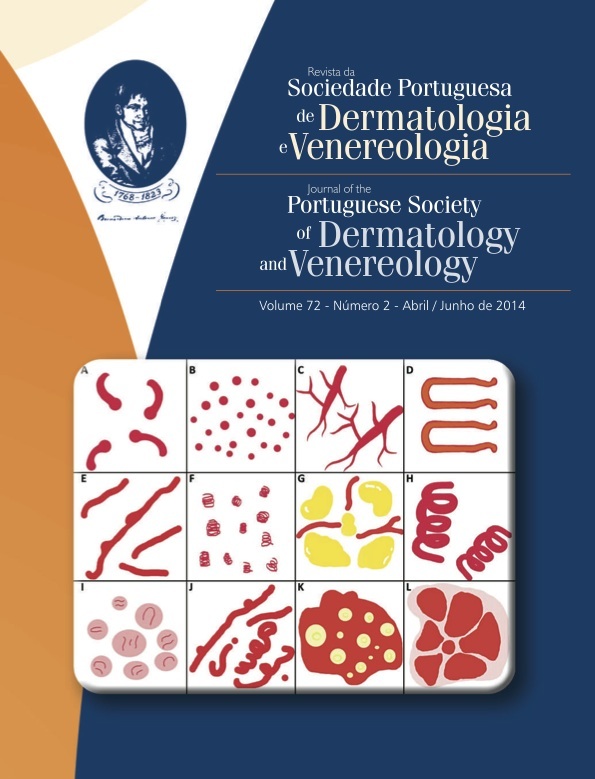
VASCULAR PATTERNS AND MORPHOLOGY IN DERMOSCOPY - PART I. BASIC PRINCIPLES
Abstract
Dermoscopy is a noninvasive, in vivo technique that increases the diagnostic accuracy in both melanocytic and nonmelanocytic skin tumors. In nonpigmented tumors allows the visualization of vascular structures not visible to the naked eye. Part I of this article discusses the dermoscopic evaluation of nonpigmented skin tumors including basic principles of specific morphologic types and architectural arrangement of vessels and the presence of additional dermoscopic clues.
Downloads
References
Zalaudek I, Jurgen Kreusch, Giacomel J, Ferrara G,
Catricalá C, Argenziano G. How to diagnose nonpigmented
skin tumors: A review of vascular structures
seen with dermoscopy. Part I Melanocytic skin
tumors. J Am Acad Dermatol. 2010; 63:361-74.
Zalaudek I, Jurgen Kreusch, Giacomel J, Ferrara G,
Catricalá C, Argenziano G. How to diagnose nonpigmented
skin tumors: A review of vascular structures
seen with dermoscopy. Part II Nonmelanocytic skin
tumors. J Am Acad Dermatol. 2010; 63:377-86.
Giacomel J, Zalaudek I. Pink Lesions. Dermatol
Clin. 2013; 31:649-78.
Marghoob AA, Liebman TN. Vascular structures. In:
Marghoob AA, Malvehy J, Braun RP, editors. Atlas
of Dermoscopy. 2nd ed. Informa healthcare; 2012.
-50.
Argenziano G. Zalaudek I, Corona R, Sera F, Cicale
L, Petrillo G, et al. Vascular Structures in Skin
Tumors. Arch Dermatol. 2004; 140: 1485-9.
All articles in this journal are Open Access under the Creative Commons Attribution-NonCommercial 4.0 International License (CC BY-NC 4.0).


