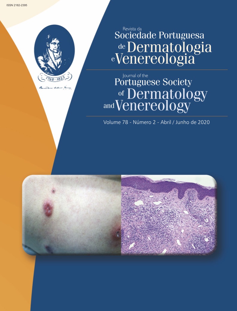
Pediatric Melanoma: Epidemiology, Pathogenesis, Diagnosis and Management
Abstract
Pediatric melanoma is the most common skin cancer in children. However, it is extremely rare this population, being even rarer in younger than 10 years of age. Its diagnosis is often difficult, due to its rarity and atypical presentations. There are three main subtypes of pediatric melanoma: Spitzoid melanoma, melanoma arising in a congenital melanocytic nevus and conventional melanoma. Congenital melanomas exist and are exceptionally rare, although they do not constitute a different subtype of melanoma. Spitzoid melanoma is the most common subtype affecting children younger than 11 years. Despite presenting with local aggressive features and frequent nodal involvement, it encompasses an excellent prognosis. The risk of malignant transformation of congenital melanocytic nevi varies widely accordingly to the projected adult size, number, and concomitant abnormalities found in the central nervous system. The surveillance and treatment of melanoma arising in a congenital melanocytic nevus is challenging, enclosing poor outcomes. In adolescents, the most common subtype is the conventional (adult-type). Contrary to the adult population, the majority of conventional pediatric melanoma arises from previous nevi but follows the general adult epidemiology and risk factors. Specific guidelines for management of pediatric melanoma do not exist and it is treated similarly to melanoma in the adult.
Downloads
References
Brecht IB, De Paoli A, Bisogno G, Orbach D, Schneider DT, Leiter U, et al. Pediatric patients with cutaneous melanoma: A European study. Pediatr Blood Cancer. 2018;65:1-8. doi:10.1002/pbc.26974
Quinlan CS, Capra M, Dempsey M. Paediatric malignant melanoma in Ireland: A population study and review of the literature. J Plast Reconstr Aesthetic Surg. 2019;72:1388-95. doi:10.1016/j.bjps.2019.03.041
Kaste SC. Imaging of pediatric cutaneous melanoma. Pediatr Radiol. 2019;49:1476-87. doi:10.1007/s00247-019-04374-9
Ipenburg NA, Lo SN, Vilain RE, Holtkamp LHJ, Wilmott JS, Nieweg OE, et al. The prognostic value of tumor mitotic rate in children and adolescents with
cutaneous melanoma: a retrospective cohort study. J Am Acad Dermatol. 2019 (in press). doi:10.1016/j.jaad.2019.10.065
Cordoro KM, Gupta D, Frieden IJ, McCalmont T, Kashani-Sabet M. Pediatric melanoma: Results of a large cohort study and proposal for modified ABCD
detection criteria for children. J Am Acad Dermatol. 2013;68:913-25. doi:10.1016/j.jaad.2012.12.953
Campbell LB, Kreicher KL, Gittleman HR, Strodtbeck K, Barnholtz-Sloan J, Bordeaux JS. Melanoma incidence in children and adolescents: Decreasing trends in the United States. J Pediatr. 2015;166:1505-13. doi:10.1016/j.jpeds.2015.02.050
Kumar RS, Messina JL, Reed DR, Sondak VK. Pediatric Melanoma and Atypical Melanocytic Neoplasms. Cancer Treat Res. 2018;167:331-69.doi:10.1007/978-3-319-78310-9
Paulson KG, Gupta D, Kim TS, Veatch JR, Byrd DR, Bhatia S, et al. Age-Specific Incidence of Melanoma in the United States. JAMA Dermatol. 2019;98109:1-8. doi:10.1001/jamadermatol.2019.3353
Merkel EA, Mohan LS, Shi K, Panah E, Zhang B, Gerami P. Paediatric melanoma: clinical update, genetic basis, and advances in diagnosis. Lancet Child
Adolesc Heal. 2019;3:646-54. doi:10.1016/S2352-4642(19)30116-6
Aldrink JH, Polites S, Lautz TB, Malek MM, Rhee D, Bruny J, et al. What’s New in Pediatric Melanoma: An Update from the APSA Cancer Committee. J Pediatr Surg. 2019 (in press). doi:10.1016/j.jpedsurg.2019.09.036
Al-Himdani S, Naderi N, Whitaker IS, Jones NW. An 18-year Study of Malignant Melanoma in Childhood and Adolescence. Plast Reconstr Surg. 2019;7:e2338. doi:10.1097/gox.0000000000002338
Bahrami A, Barnhill RL. Pathology and Genomics of Pediatric Melanoma: A Critical Re-examination and New Insights. Pediatr Blood Cancer. 2018;65. doi:10.1016/j.physbeh.2017.03.040
Stefanaki C, Chardalias L, Soura E, Katsarou A, Stratigos S. Pediatric melanoma. J Eur Acad Dermatol Venereol. 2017;31:1604-15. doi:10.1111/ijlh.12426
Bartenstein DW, Fisher JM, Stamoulis C, Weldon C, Huang JT, Gellis SE, et al. Clinical features and outcomes of spitzoid proliferations in children and adolescents. Br J Dermatol. 2019;181:366-72. doi:10.1111/bjd.17450
Tas F, Erturk K. Spitzoid cutaneous melanoma is associated with favorable clinicopathological factors and outcome. J Cosmet Dermatol. 2019 (in press). doi:10.1111/jocd.12958
Gerami P, Scolyer RA, Xu X, Elder DE, Abraham RM, Fullen D, et al. Risk assessment for atypical spitzoid melanocytic neoplasms using FISH to identify chromosomal copy number aberrations. Am J Surg Pathol. 2013;37:676-84. doi:10.1097/PAS.0b013e3182753de6
Lee S, Barnhill RL, Dummer R, Dalton J, Wu J, Pappo A, et al. TERT Promoter Mutations Are Predictive of Aggressive Clinical Behavior in Patients with Spitzoid Melanocytic Neoplasms. Sci Rep. 2015;5:1-7. doi:10.1038/srep11200
Lee CY, Sholl LM, Zhang B, Merkel EA, Amin SM, Guitart J,et al. Atypical Spitzoid Neoplasms in Childhood. Am J Dermatopathol. 2017;39:181-6. doi:10.1097/dad.0000000000000629
Lallas A, Apalla Z, Ioannides D, Lazaridou E, Kyrgidis A, Broganelli P, et al. Update on dermoscopy of Spitz/Reed naevi and management guidelines by the International Dermoscopy Society. Br J Dermatol. 2017;177:645-55. doi:10.1111/ijlh.12426
Moscarella E, Lallas A, Kyrgidis A, Ferrara G, Longo C, Scalvenzi M, et al. Clinical and dermoscopic features of Atypical Spitz Tumors: a multi-center, retrospective, case-control study. J Am Acad Dermatol. 2015;73:777-84. doi:10.1016/j.jaad.2015.08.018.Clinical
Lazova R, Seeley EH, Kutzner H, Scolyer RA, Scott G, Cerroni L,et al. Imaging mass spectrometry assists in the classification of diagnostically challenging atypical Spitzoid neoplasms. J Am Acad Dermatol. 2016;75:1176-1186.e4. doi:10.1016/j.jaad.2016.07.007
Lallas A, Kyrgidis A, Ferrara G, Atypical Spitz tumours and sentinel lymph node biopsy: A systematic review et al. Atypical Spitz tumours and sentinel lymph node biopsy: A systematic review. Lancet Oncol. 2014;15:e178-e183. doi:10.1016/S1470-2045(13)70608-9
Kinsler VA, O’Hare P, Bulstrode N, Calonje JE, Chong WK, Hargrave D, et al. Melanoma in congenital melanocytic naevi. Br J Dermatol. 2017;176:1131-43. doi:10.1111/bjd.15301
Viana ACL, Goulart EM, Gontijo B, Bittencourt FV. A prospective study of patients with large congenital melanocytic nevi and the risk of melanoma. An Bras Dermatol. 2017;92:200-5. doi:10.1590/abd1806-4841.20175176
Polubothu SD, Mcguire N, Baird W, Baird W, Bulstrode N, Chalker J, et al. Does the gene matter ? Genotype – phenotype and genotype – outcome associations in congenital melanocytic naevi. Br J Dermatol. 2020; ;182:434-43. doi:10.1111/bjd.18106
Price HN. Congenital melanocytic nevi: Update in genetics and management. Curr Opin Pediatr. 2016;28:476-82. doi:10.1097/MOP.0000000000000384
Neuhold JC, Friesenhahn J, Gerdes N, Krengel S. Case reports of fatal or metastasizing melanoma in children and adolescents: A systematic analysis of the literature. Pediatr Dermatol. 2015;32:13-22. doi:10.1111/pde.12400
Ott H, Krengel S, Beck O, Böhler K, Böttcher-Haberzeth S, Cangir Ö, et al. Multidisciplinary long-term care and modern surgical treatment of congenital melanocytic nevi – recommendations by the CMN surgery network. J Dtsch Dermatol Ges. 2019;17:1005-16. doi: 10.1111/ddg.13951.
Wisell J. Proliferative Nodules Arising Within Congenital Melanocytic Nevi: A Histologic, Immunohistochemical, and Molecular Analyses of 43 Cases. Yearb Pathol Lab Med. 2012;2012:74-5. doi:10.1016/j.ypat.2011.11.123
Friedman EB, Scolyer RA, Thompson JF. Management of pigmented skin lesions in childhood and adolescence. Aust J Gen Pract. 2019;48:539-44. doi:10.31128/AJGP-04-19-48951
Waelchli R, Aylett SE, Atherton D, Thompson DJ, Chong WK, Kinsler VA. Classification of neurological abnormalities in children with congenital melanocytic naevus syndrome identifies magnetic resonance imaging as the best predictor of clinical outcome. Br J Dermatol. 2015;173:739-50. doi:10.1111/bjd.13898
Aoude LG, Wadt KAW, Pritchard AL, Hayward NK. Genetics of familial melanoma: 20 years after CDKN2A. Pigment Cell Melanoma Res. 2015;28:148-60.
doi:10.1111/pcmr.12333
Landi MT, Bauer J, Pfeiffer RM, Elder DE, Hulley B, Minghetti P,et al. MC1R germline variants confer risk for BRAF-mutant melanoma. Science. 2006;313:521-2. doi:10.1126/science.1127515
Richardson SK, Tannous ZS, Mihm MC. Congenital and infantile melanoma: Review of the literature and report of an uncommon variant, pigment-synthesizing melanoma. J Am Acad Dermatol. 2002;47:77-90. doi:10.1067/mjd.2002.120602
Kim J, Sun Z, Gulack BC, Adam MA, Mosca PJ, Rice HE, et al. Sentinel lymph node biopsy is a prognostic measure in pediatric melanoma. J Pediatr Surg. 2016;51:986-90. doi:10.1016/j.jpedsurg.2016.02.067
Wong SL, Faries MB, Kennedy EB, Agarwala SS, Akhurst TJ, Ariyan C, et al. Sentinel lymph node biopsy and management of regional lymph nodes in Melanoma: American society of clinical oncology and society of surgical oncology clinical practice guideline update. J Clin Oncol. 2018;36:399-413. doi:10.1200/JCO.2017.75.7724
Navid F, Furman WL, Fleming M, Rao BN, Kovach S, Billups CA, et al. The feasibility of adjuvant interferon α-2b in children with high-risk melanoma. Cancer. 2005;103:780-7. doi:10.1002/cncr.20860
Kinsler VA, O’Hare P, Jacques T, Hargrave D, Slater O. MEK inhibition appears to improve symptom control in primary NRAS-driven CNS melanoma in children. Br J Cancer. 2017;116:990-3. doi:10.1038/bjc.2017.49
Murphy BM, Burd CE. Can Combination MEK and Akt Inhibition Slay the Giant Congenital Nevus? J Invest Dermatol. 2019;139:1857-9. doi:10.1016/j.
jid.2019.04.009
Rouillé T, Aractingi S, Kadlub N, Fraitag S, How-Kit A, Daunay A, et al.. Local Inhibition of MEK/Akt Prevents Cellular Growth in Human Congenital Melanocytic Nevi. J Invest Dermatol. 2019;139(9):2004-2015.e13. doi:10.1016/j.jid.2019.03.1156
Hodi FS, Corless CL, Giobbie-Hurder A, Fletcher JA, Zhu M, Marino-Enriquez A, et al. Imatinib for melanomas harboring mutationally activated or amplified kit arising on mucosal, acral, and chronically sun-damaged skin. J Clin Oncol. 2013;31;31:3182-90. doi:10.1200/JCO.2012.47.7836
Copyright (c) 2020 Journal of the Portuguese Society of Dermatology and Venereology

This work is licensed under a Creative Commons Attribution-NonCommercial 4.0 International License.
All articles in this journal are Open Access under the Creative Commons Attribution-NonCommercial 4.0 International License (CC BY-NC 4.0).


