New World Leishmaniasis: The Role of Confocal Microscopy in Diagnosis and Follow-up - Tropical Dermatology
Resumo
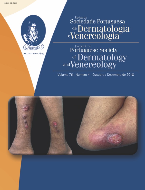
Cutaneous leishmaniasis may mimic other infections in overlapping endemic areas and timely treatment prevents dissemination of the parasite. The required histopathological and microbiological examinations are not always available, and can only give a deferred confirmation of the diagnosis. In contrast, reflectance confocal microscopy (RCM) allows real-time visualization till the level of papillary dermis. A 59-year-old Brazilian male presented with ulcerated plaques and tumors on the extremities. The clinical differential diagnosis included leishmaniasis and other infections with lymphocutaneous pattern of dissemination. RCM showed the characteristic picture of «eggs in a bird’s nest» which has been described in cutaneous leishmaniasis. The diagnosis of leishmaniasis was later confirmed by skin biopsy, in which Leishmania guyanensis was identified by parasitological examination. After treatment with liposomal amphotericin B, reassessment with RCM corroborated the clinical cure, showing an «empty nest» picture. In conclusion, RCM noninvasively provides useful information for diagnosis and follow-up of cutaneous leishmaniasis.
Downloads
Referências
Handler MZ, Patel PA, Kapila R, Al-Qubati Y, Schwartz
RA. Cutaneous and mucocutaneous leishmaniasis: Differential
diagnosis, diagnosis, histopathology, and
management. J Am Acad Dermatol. 2015;73:911-26.
doi: 10.1016/j.jaad.2014.09.014
Alvar J, Vélez I, Bern C, Herrero M, Desjeux P, Cano
J, et al. WHO Leishmaniasis Control Team: Leishmaniasis
worldwide and global estimates of its incidence.
PLoS One. 2012;7:e35671. doi: 10.1371/journal.
pone.0035671.
World Health Organization. Control of the leishmaniases.
World Heal Organ Tech Rep Ser. 2010;949:1-
Buljan M, Zalaudek I, Massone C, Hofmann-Wellenhof
R, Fink-Puches R, Arzberger E. Dermoscopy and reflectance
confocal microscopy in cutaneous leishmaniasis
on the face. Australas J Dermatol. 2016;57:316-8.
doi: 10.1111/ajd.12404.
Alarcon I, Carrera C, Puig S, Malvehy J. In vivo
confocal microscopy features of cutaneous leishmaniasis.
Dermatology. 2014;228:121-4. doi:
1159/000357525.
Goihman-Yahr M. American mucocutaneous leishmaniasis.
Dermatol Clin. 1994;12:703-12.
Vega-Lopez F. Diagnosis of cutaneous leishmaniasis.
Curr Opin Infect Dis. 2003;16:97-101.
Al-Hucheimi S, Sultan B, Al-Dhalimi M. A comparative
study of the diagnosis of Old World cutaneous leishmaniasis
in Iraq by polymerase chain reaction and
microbiologic and histopathologic methods. Int J Dermatol.
;48:404-8. doi: 10.1159/000357525.
Skoryna-Karcz B, Chojecka-Adamska A, Adamski Z.
The case of cutaneous leishmaniasis - diagnostic difficulties.
Przegl Dermatol. 1994;81:544-7.
Kalter D. Laboratory tests for the diagnosis and evaluation
of leishmaniasis. Dermatol Clin. 1994;12:37-
Practical guide for specimen collection and reference
diagnosis of leishmaniasis [Internet]. US Centers for
Disease Control and Prevention Website. 2014 [cited
Aug 20]. Available from: https://www.cdc.gov/
parasites/leishmaniasis/resources/pdf/cdc_diagnosis_
guide_leishmaniasis.pdf
Llambrich A, Zaballos P, Terrasa F, Torne I, Puig S, Malvehy
J. Dermoscopy of cutaneous leishmaniasis. Br J
Dermatol. 2009;160:756-61. doi: 10.1111/j.1365-
-2133.2008.08986.x.
Yucel A, Gunasti S, Denli Y, Uzun S. Cutaneous
leishmaniasis: new dermoscopic findings. Int J Dermatol.
;52:831-7. doi: 10.1111/j.1365-
-4632.2012.05815.x.
Rajadhyaksha M, Gonza S, Zavislan JM, Anderson RR,
Webb RH. In Vivo Confocal Scanning Laser Microscopy
of Human Skin II : Advances in Instrumentation and
Comparison With. J Invest Dermatol. 1999;113:293-
Cinotti E, Perrot JL, Labeille B, Cambazard F. Reflectance
confocal microscopy for cutaneous infections
and infestations. J Eur Acad Dermatol Venereol. 2016;30:754-63. doi: 10.1111/jdv.13254.
Cinotti E, Perrot JL, Labeille B, Vercherin P, Chol C,
Besson E, et al. Reflectance confocal microscopy for
quantification of Sarcoptes scabiei in Norwegian scabies.
J Eur Acad Dermatol Venereol. 2013;27:e176-8.
doi: 10.1111/j.1468-3083.2012.04555.x.
Slutsky JB, Rabinovitz H, Grichnik JM, Marghoob
AA. Reflectance confocal microscopic features of
dermatophytes, scabies, and Demodex. Arch Dermatol.
;147:1008. doi: 10.1001/archdermatol.
193.
Goldgeier M, Alessi C, Muhlbauer J. Immediate noninvasive
diagnosis of herpesvirus by confocal scanning
laser microscopy. J Am Acad Dermatol. 2002;46:783-5.
Todos os artigos desta revista são de acesso aberto sob a licença internacional Creative Commons Attribution-NonCommercial 4.0 (CC BY-NC 4.0).


