
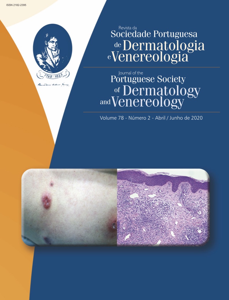
Dermatoses com Alterações Histológicas Mínimas: Tornar o Invisível Visível
Resumo
As biópsias cutâneas continuam a ser uma ferramenta indispensável no auxílio de dermatologistas para um diagnóstico e tratamento precisos. Contudo, algumas doenças dermatológicas clinicamente evidentes mostram imagens histológicas normais, quando examinadas após preparação com hematoxilina-eosina (H&E). No sentido de estabelecer um diagnóstico correcto, é essencial a correlação clínico-patológica ou executar outras investigações adicionais, como colorações especiais e técnicas de imuno-histoquímica. Neste artigo, são discutidas as mais relevantes destas dermatoses “invisíveis” em H&E, incluindo a estratégia de abordagem nestes casos.
Downloads
Referências
Brownstein MH, Rabinowitz A. The invisible dermatoses. J Am Acad Dermatol. 1983;8(4):579-588.
Requena L, Kutzner H. [Invisible dermatosis]. Pathologe. 2002;23(1):54-64.
Tomasini C. Invisible dermatoses from the perspective of the dermatopathologist: new observations. G Ital di dermatologia e Venereol organo Uff Soc Ital di dermatologia e Sifilogr. 2017;152(5):500-515.
Requena L, Sánchez Yus E. Invisible dermatoses. Additional findings. Int J Dermatol.
Mysore V. Invisible dermatoses. Indian J Dermatol Venereol Leprol. 2010;76(3):239-248.
Carlotti A. [Invisible dermatoses]. Ann Dermatol Venereol. 2009;136(2):152-159.
Tomasini C, Michelerio A. Invisible dermatoses: clues and pitfalls to diagnosis. Diagnostic Histopathol. 2018;24(8):320-337.
Cardoso JC, Veraitch O, Gianotti R, et al. “Hints” in the horn: diagnostic clues in the stratum corneum. J Cutan Pathol. 2017;44(3):256-278.
Park YW, Kim DY, Yoon SY, et al. “Clues” for the histological diagnosis of tinea: how reliable are they? Ann Dermatol. 2014;26(2):286-288.
Fernandez N, Torres A, Ackerman AB. Pathologic Findings in Human Scabies. Arch Dermatol. 1977;113(3):320-324.
Kim YC, Kim YJ, Kang HY, Sohn S, Lee E-S. Histopathologic features in vitiligo. Am J Dermatopathol. 2008;30(2):112-116.
Alhumidi A, Alshamlan N, Alfaraidi M, Mohajer K. Invisible dermatosis, diagnostic discrepancy between the general pathologist and dermatopathologist. J Cutan Pathol. 2019;46(12):905-912.
Johnston RB. 8 - Vasculopathic Reaction Pattern. In: Johnston RB, ed. Weedon’s Skin Pathology Essentials (Second Edition). Second Edi. Elsevier; 2017:163-194.
Antia C, Baquerizo K, Korman A, Bernstein JA, Alikhan A. Urticaria: A comprehensive review: Epidemiology, diagnosis, and work-up. J Am Acad Dermatol. 2018;79(4):599-614.
Severino M, Chandesris M-O, Barete S, et al. Telangiectasia macularis eruptiva perstans (TMEP): A form of cutaneous mastocytosis with potential systemic involvement. J Am Acad Dermatol. 2016;74(5):885-91.e1
Brownstein MH, Hashimoto K. Macular amyloidosis. Arch Dermatol. 1972;106(1):50-57.
Walters R, Pulitzer M, Kamino H. Elastic fiber pattern in scleroderma/morphea. J Cutan Pathol. 2009;36(9):952-957.
Direitos de Autor (c) 2020 Revista da Sociedade Portuguesa de Dermatologia e Venereologia

This work is licensed under a Creative Commons Attribution-NonCommercial 4.0 International License.
Todos os artigos desta revista são de acesso aberto sob a licença internacional Creative Commons Attribution-NonCommercial 4.0 (CC BY-NC 4.0).


