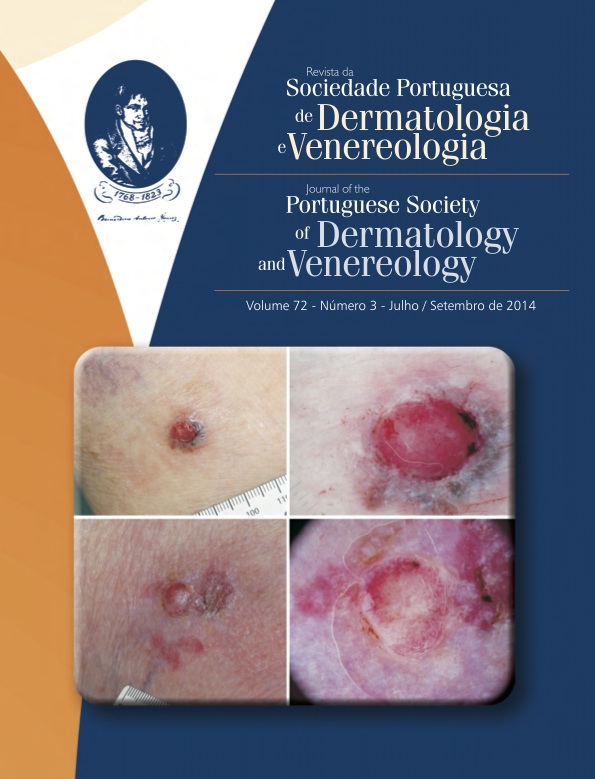
VASCULAR PATTERNS AND MORPHOLOGY IN DERMOSCOPY - PART II. CLINICAL PRACTICE
Keywords:
actinic, Carcinoma, basal cell, intradermal, Dermoscopy, Melanoma, Skin neoplasms, Keratosis, seborrheic, squamous cell, Nevus, Epithelioid and spindle cell
Abstract
Dermoscopy is a noninvasive, in vivo technique that increases the diagnostic accuracy in both melanocytic and nonmelanocytic skin tumors. In nonpigmented tumors it allows the visualization of vascular structures not visible to the naked eye. Part II of this article discusses clinical applications of dermoscopy in non-pigmented tumoral skin lesions.
Downloads
Download data is not yet available.
Published
2014-10-12
How to Cite
Laureano, A., Fernandes, C., & Cardoso, J. (2014). VASCULAR PATTERNS AND MORPHOLOGY IN DERMOSCOPY - PART II. CLINICAL PRACTICE. Journal of the Portuguese Society of Dermatology and Venereology, 72(3), 307-324. https://doi.org/10.29021/spdv.72.3.273
Section
Continuous Medical Education
All articles in this journal are Open Access under the Creative Commons Attribution-NonCommercial 4.0 International License (CC BY-NC 4.0).


