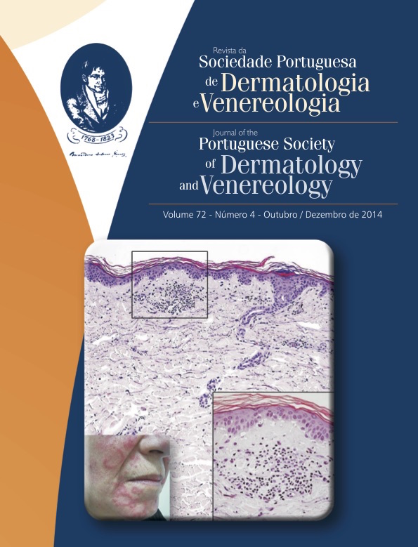
LÚPUS ERITEMATOSO CUTÂNEO - CORRELAÇÃO CLÍNICOPATOLÓGICA DE MANIFESTAÇÕES CUTÂNEAS ESPECÍFICAS
Resumo
Introdução: O lúpus eritematoso (LE) é uma patologia multiorgânica autoimune que afeta sobretudo mulheres em idade fértil. A pele é o segundo órgão mais frequentemente atingido, depois das articulações, e pode preceder o envolvimento sistémico.
Objetivos: Apresentar aspetos clínicos que evocam a hipótese de manifestações cutâneas específicas de LE e achados histopatológicos que podem contribuir para fundamentar o diagnóstico.
Métodos: Estudo de casos com manifestações cutâneas sugestivas de LE, nos quais a correlação clínico-histopatológica possibilitou o diagnóstico de diferentes subtipos de LE.
Resultados: A correlação das alterações clínicas e histopatológicas, no contexto analítico apropriado, permitiu identificar casos concretos de manifestações cutâneas específicas de LE: 1) agudo; 2) subagudo (formas anular e papular-escamosa); 2.1) subagudo induzido por fármacos; 3) crónico, nomeadamente 3.1) discóide clássico (face e couro cabeludo); 3.2) túmido; 3.3) paniculite.
Descreve-se, adicionalmente, os achados clínicos e histopatológicos expectáveis em outras manifestações cutâneas específicas de LE.
Conclusão: O LE exibe grande variabilidade de manifestações cutâneas. A histopatologia é essencial para o diagnóstico, sendo muito importante a preservação e manuseamento apropriado do material de biópsia. A imunofluorescência direta é útil para melhorar a acuidade diagnóstica. O diagnóstico preciso, baseado na correlação clínico-patológica sistemática e integrado no contexto clínico-analítico adequado, é fundamental para o prognóstico e orientação terapêutica dos doentes.
Downloads
Referências
Tsokos GC. Systemic lupus erythematosus. N Engl J Med. 2011; 365:2110-21.
Petri M, Orbai AM, Alarcon GS, Gordon C, Merrill JT, Fortin PR, et al. Derivation and validation of the systemic lupus international collaborating clinics classification criteria for systemic lupus erythematosus. Arthritis Rheum. 2012; 64:2677-86.
Albrecht J, Berlin JA, Braverman IM, Callen JP, Connolly MK, Costner MI, et al. Dermatology position
paper on the revision of the 1982 acr criteria for systemic lupus erythematosus. Lupus. 2004; 13:839-49.
Obermoser G, Sontheimer RD, Zelger B. Overview of common, rare and atypical manifestations of cutaneous lupus erythematosus and histopathological correlates. Lupus. 2010; 19:1050-70.
Shatley MJ, Walker BL, McMurray RW. Lues and lupus: Syphilis mimicking systemic lupus erythematosus (sle). Lupus. 2001; 10:299-303.
Costner MI, Sontheimer RD. Lupus erythematosus. In: Wolff K, Goldsmith LA, Katz SI, Gilchrest BA, Paller AS, Leffell DJ, editors Fitzpatrick´s Dermatology in General Medicine. 8th ed. New York, NY: McGraw Hill; 2012 p.1909-1926.
Gilliam JN, Sontheimer RD. Distinctive cutaneous subsets in the spectrum of lupus erythematosus. J
Am Acad Dermatol. 1981; 4:471-5.
Crowson AN, Magro C. The cutaneous pathology of lupus erythematosus: A review. J Cutan Pathol. 2001; 28:1-23.
Rothfield N, Sontheimer RD, Bernstein M. Lupus erythematosus: Systemic and cutaneous manifestations. Clin Dermatol. 2006; 24:348-62.
Sepehr A, Wenson S, Tahan SR. Histopathologic manifestations of systemic diseases: The example of cutaneous lupus erythematosus. J Cutan Pathol. 2010;37 Suppl 1:112-24.
Crowson AN, Mihm MC, Jr., Magro CM. Cutaneous vasculitis: A review. J Cutan Pathol. 2003; 30:161-73.
Lipsker D, Saurat JH. Neutrophilic cutaneous lupus erythematosus. At the edge between innate and acquired immunity? Dermatology. 2008; 216:283-6.
Yell JA, Allen J, Wojnarowska F, Kirtschig G, Burge SM. Bullous systemic lupus erythematosus: Revised criteria for diagnosis. Br J Dermatol. 1995; 132:921-8.
Rappersberger K, Tschachler E, Tani M, Wolff K. Bullous disease in systemic lupus erythematosus. J Am Acad Dermatol. 1989; 21:745-52.
Sontheimer RD, Thomas JR, Gilliam JN. Subacute cutaneous lupus erythematosus: A cutaneous marker
for a distinct lupus erythematosus subset. Arch Dermatol. 1979; 115:1409-15.
Crowson AN, Magro CM, Mihm MC, Jr. Interface dermatitis. Arch Pathol Lab Med. 2008; 132:652-66.
Vedove CD, Del Giglio M, Schena D, Girolomoni G. Drug-induced lupus erythematosus. Arch Dermatol Res. 2009; 301:99-105.
Marzano AV, Vezzoli P, Crosti C. Drug-induced lupus: An update on its dermatologic aspects. Lupus. 2009; 18:935-40.
Gronhagen CM, Fored CM, Linder M, Granath F, Nyberg F. Subacute cutaneous lupus erythematosus and its association with drugs: A population-based matched case-control study of 234 patients in sweden. Br J Dermatol. 2012; 167:296-305.
Crowson AN, Magro CM. Recent advances in the pathology of cutaneous drug eruptions. Dermatol Clin. 1999; 17:537-60.
Al-Refu K, Goodfield M. Scar classification in cutaneous lupus erythematosus: Morphological description. Br J Dermatol. 2009; 161:1052-8.
Perniciaro C, Randle HW, Perry HO. Hypertrophic discoid lupus erythematosus resembling squamous cell carcinoma. Dermatol Surg. 1995; 21:255-7.
Nico MM, Vilela MA, Rivitti EA, Lourenco SV. Oral lesions in lupus erythematosus: Correlation with cutaneous lesions. Eur J Dermatol. 2008; 18:376-81.
Orteu CH, Buchanan JA, Hutchison I, Leigh IM, Bull RH. Systemic lupus erythematosus presenting with oral mucosal lesions: Easily missed? Br J Dermatol. 2001; 144:1219-23.
Kuhn A, Richter-Hintz D, Oslislo C, Ruzicka T, Megahed M, Lehmann P. Lupus erythematosus tumidus - a neglected subset of cutaneous lupus erythematosus: Report of 40 cases. Arch Dermatol. 2000; 136:1033-41.
Kuhn A, Sonntag M, Ruzicka T, Lehmann P, Megahed M. Histopathologic findings in lupus erythematosus tumidus: Review of 80 patients. J Am Acad Dermatol. 2003; 48:901-8.
Schmitt V, Meuth AM, Amler S, Kuehn E, Haust M, Messer G, et al. Lupus erythematosus tumidus is a separate subtype of cutaneous lupus erythematosus. Br J Dermatol. 2010; 162:64-73.
Arai S, Katsuoka K. Clinical entity of lupus erythematosus panniculitis/lupus erythematosus profundus.
Autoimmun Rev. 2009; 8:449-52.
Massone C, Kodama K, Salmhofer W, Abe R, Shimizu H, Parodi A, et al. Lupus erythematosus panniculitis (lupus profundus): Clinical, histopathological, and molecular analysis of nine cases. J Cutan Pathol. 2005; 32:396-404.
Chopra R, Chhabra S, Thami GP, Punia RP. Panniculitis: Clinical overlap and the significance of biopsy findings. J Cutan Pathol. 2010; 37:49-58.
Sontheimer RD. Skin manifestations of systemic autoimmune connective tissue disease: Diagnostics and therapeutics. Best Pract Res Clin Rheumatol. 2004; 18:429-62.
Magro CM, Crowson AN. The cutaneous pathology associated with seropositivity for antibodies to ssa (ro): A clinicopathologic study of 23 adult patients without subacute cutaneous lupus erythematosus. Am J Dermatopathol. 1999; 21:129-37.
Hedrich CM, Fiebig B, Hauck FH, Sallmann S, Hahn G, Pfeiffer C, et al. Chilblain lupus erythematosus-a review of literature. Clin Rheumatol. 2008; 27:1341.
Crowson AN, Magro CM. Idiopathic perniosis and its mimics: A clinical and histological study of 38 cases. Hum Pathol. 1997; 28:478-84.
Provost TT, Andres G, Maddison PJ, Reichlin M. Lupus band test in untreated sle patients: Correlation
of immunoglobulin deposition in the skin of the extensor forearm with clinical renal disease and serological abnormalities. J Invest Dermatol. 1980; 74:407-12.
Magro CM, Dyrsen ME. The use of c3d and c4d immunohistochemistry on formalin-fixed tissue as a diagnostic adjunct in the assessment of inflammatory skin disease. J Am Acad Dermatol. 2008; 59:822-33.
Weigand DA. Lupus band test: Anatomic regional variations in discoid lupus erythematosus. J Am Acad Dermatol. 1986; 14:426-8.
Todos os artigos desta revista são de acesso aberto sob a licença internacional Creative Commons Attribution-NonCommercial 4.0 (CC BY-NC 4.0).


