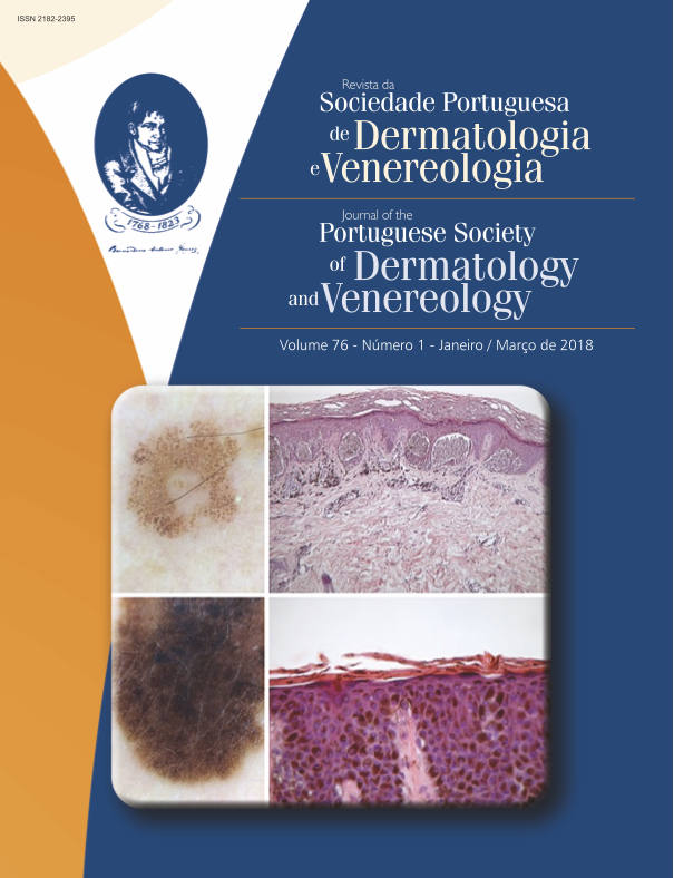
Diagnostic Accuracy of Dermoscopy in Melanocytic Lesions: Retrospective Study with Histological Correlation
Abstract
Introduction: Dermoscopy is an in vivo, non-invasive technique that is widely used as a complementary method for the study of pigmented skin lesions and enables the early diagnosis of cutaneous melanoma. The purpose of the present study is to evaluate the diagnostic accuracy of dermoscopy and to extend the dermoscopy-histopatholoy correlations.
Methods: Retrospective study using the database of the Dermoscopy and Pigmented Lesions Clinic and the database of the Histology of the Dermatology Department of CHUC. Each melanocytic lesion was evaluated according to multiple dermoscopic and histopathological parameters. The agreement between diagnoses and between the evaluated parameters was determined.
Results: The malignant/benign ratio was 1:9.2. The agreement between the dermoscopic and histologic diagnoses was fair. The agreement for melanoma was excellent and dermoscopy showed 92.9% sensitivity and 96.9% specificity for this diagnosis. There was agreement between a reticular pattern and the presence of bridging of rete ridges and cytological atypia; globular pattern and the presence of nests; atypical network and presence of fibrosis and cytological atypia; blotches and pigmented parakeratosis; blue-whitish veil and fibrosis.
Conclusion: In spite of some limitations and unexpected findings, many of the results of the present study are concordant with the literature, showing a high sensitivity and specificity of dermoscopy in the diagnosis of melanoma, which results in improvement in early diagnosis without increasing the total number of excisions of pigmented lesions.
Downloads
References
Campos-do-Carmo G, Ramos-e-Silva M. Dermoscopy: basic concepts. Int J Dermatol. 2008;47:712-9.
Roldán-Marín R, Puig S, Malvehy J. Dermoscopic criteria and melanocytic lesions. G Ital Dermatol Venereol. 2012;147:149-59.
Menezes N. Dermatoscopia de lesões pigmentadas. Trab Soc Port Dermatol Venereol. 2011;69:33-48.
Ungureanu L, Şenila S, Danescu S, Rogojan L, Cosgarea R. Correlation of dermatoscopy with the histopathological changes in the diagnosis of thin melanoma. Rom J Morphol Embryol. 2013;54:315-20.
Massi D, Giorgi VD, Soyer HP. Histopathologic correlates of dermoscopic criteria. Dermatol Clin. 2001;19:259-68.
Morales-Callaghan AM, Castrodeza-Sanz J, Martínez-García G, Peral-Martínez I, Miranda-Romero A. Estudio de correlación clínica , dermatoscópica e histopatológica de nevus melanocíticos atípicos. Actas Dermosifiogr. 2008;99:380-9.
Álvarez CC, Zaballos P, Puig S, Malvehy J, Mascaró-Galy JM, Palou J. Correlación histológica en dermatoscopia; lesiones melanocíticas y no melanocíticas. Criterios dermatoscópicos de nevus melanocíticos. Med Cutan Ibero Lat Am. 2004;32:47-60.
Ferrara G, Argenziano G, Soyer HP, Staibano S, Ruocco E, De Rosa G. Dermoscopic-pathologic correlation: an atlas of 15 cases. Clin Dermatol. 2002;20:228-35.
Bauer J, Metzler G, Rassner G, Garbe C, Blum A. Dermatoscopy turns histopathologist’s attention to the suspicious area in melanocytic lesions. Arch Dermatol. 2001;137:1338-40.
Antonio JR, D’Avila S, Trídico L, Soubhia R, Caldas A, Alves F. Correlation between dermoscopic and histopathological diagnoses of atypical nevi in a dermatology outpatient clinic of the Medical School of São José do Rio Preto, SP, Brazil. An Bras Dermatol. 2013; 88:199-203.
Kittler H, Pehamberger H, Wolff K, Binder M. Diagnostic accuracy of dermoscopy. Lancet Oncol. 2002;3:159-65.
International Agency for Research on Cancer. Country: Portugal. IARC; 2012 [accessed 10 feb 2016] Available from: http://eco.iarc.fr/eucan/Country.aspx?ISOCountryCd=620.
Piliouras P, Gilmore S, Wurm EM, Soyer HP, Zalaudek I. New insights in naevogenesis: number, distribution and dermoscopic patterns of naevi in the elderly. Australas J Dermatol. 2011;52:254-8.
Landis JR, Koch GC. The measurement of observer agreement of categorical data. Biometrics.
;33:159-74.
Di Stefani A, Massone C, Soyer HP, Zalaudek I, Argenziano G, Arzberger E, et al. Benign dermoscopic features in melanoma. J Eur Acad Dermatology Venereol. 2014;28:799-804.
Argenziano G, Soyer HP, Chimenti S, Talamini R, Corona R, Sera F, et al. Dermoscopy of pigmented skin lesions: Results of a consensus meeting via the internet. J Am Acad Dermatol. 2003;48:679-93.
Merkel EA, Amin SM, Lee CY, Rademaker AW, Yazdan P, Martini MC, et al. The utility of dermoscopy-guided histologic sectioning for the diagnosis of melanocytic lesions: A case-control study. J Am Acad Dermatol. 2016;74:1-7.
Scope A, Busam KJ, Malvehy J, Puig S, McClain SA, Braun RP, et al. Ex vivo dermoscopy of melanocytic tumors: time for dermatopathologists to learn dermoscopy. Arch Dermatol. 2007;143:1548-52.
Carli P, De Giorgi V, Crocetti E, Mannone F, Massi D, Chiarugi A, et al. Improvement of malignant/benign ratio in excised melanocytic lesions in the “dermoscopy era”: A retrospective study 1997-2001. Br J Dermatol. 2004;150:687-92.
Haspeslagh M, Degryse N, De Wispelaere I. Routine use of ex vivo dermoscopy with “derm dotting” in dermatopathology. Am J Dermatopathol. 2013;35:867-9.
Braun RP, Kaya G, Masouyé I, Krischer J, Saurat J-H. Histopathologic Correlation on Dermoscopy: A Micropunch Technique. Arch Dermatol. 2003;139:349-51.
Haspeslagh M, Vossaert K, Lanssens S, Noë M, Hoorens I, Chevolet I, et al. Comparison of ex vivo and in vivo dermoscopy in dermatopathologic evaluation of skin tumors. JAMA Dermatol. 2016;152:1-6.
Cabete J, Lencastre A, João A. Combined use of ex vivo dermoscopy and histopathology for the diagnosis of melanocytic tumors. Am J Dermatopathol. 2016;38:189-93.
All articles in this journal are Open Access under the Creative Commons Attribution-NonCommercial 4.0 International License (CC BY-NC 4.0).


